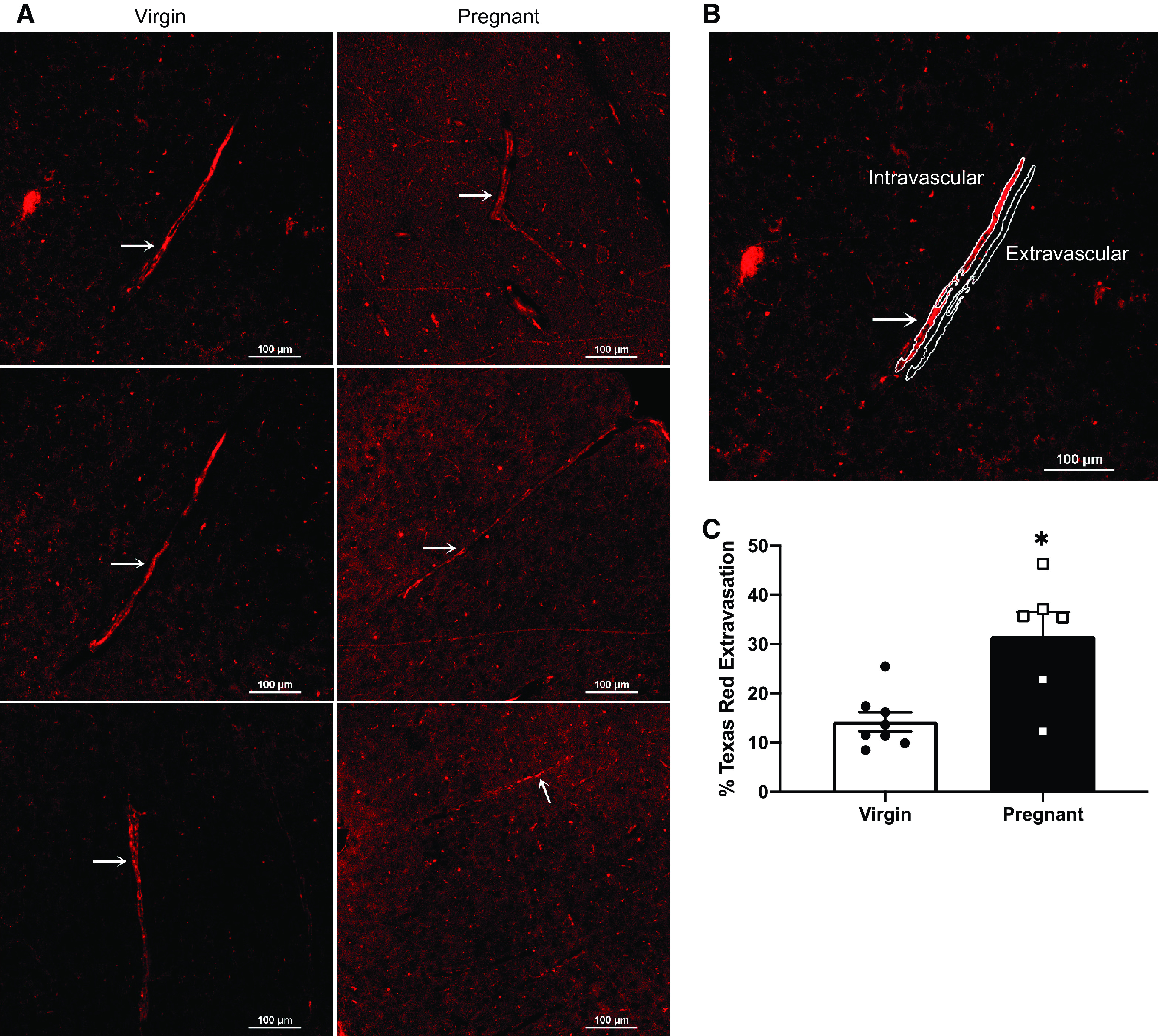
Figure 3.

Cerebrovascular extravasation of Texas Red dextran: representative images (A and B) and quantification (C) of pregnant Dahl SS/jr rats displaying an increased extravasation of 3 kDa Texas Red dextran compared with virgin littermates. Arrow denotes the vessel analyzed in the representative images. *P < 0.05; virgin n = 6 rats, pregnant n = 8 rats.
