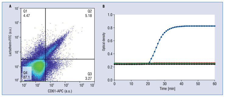
Figure 5.
Proposed procoagulant activity of platelet extracellular vesicles (PEVs) in plasma. Fibrin generation test was performed in platelet-depleted, but PEVs-containing plasma stimulated with adenosine diphosphate (ADP). A part of PEVs expose phosphatidylserine (CD61+/PS+) (A). When the plasma clots, the optical density increases (B). ADP-stimulated plasma containing phosphatidylserine (PS)-exposing extracellular vesicles (EVs) did not clot (red). Upon the addition of recombinant human tissue factor (TF) to this plasma, clotting was triggered as measured by fibrin generation (blue). The extent of clotting was not affected by the presence of the P2Y1 and P2Y12 receptor antagonists (data not shown). Covering PS-exposing EVs with an excess of lactadherin, or inhibiting the TF-initiated extrinsic coagulation with an anti-factor VIIa antibody, both inhibited plasma clotting (black and green, respectively). Hence, both PS-containing EVs and TF are indispensable for propagation of coagulation.

