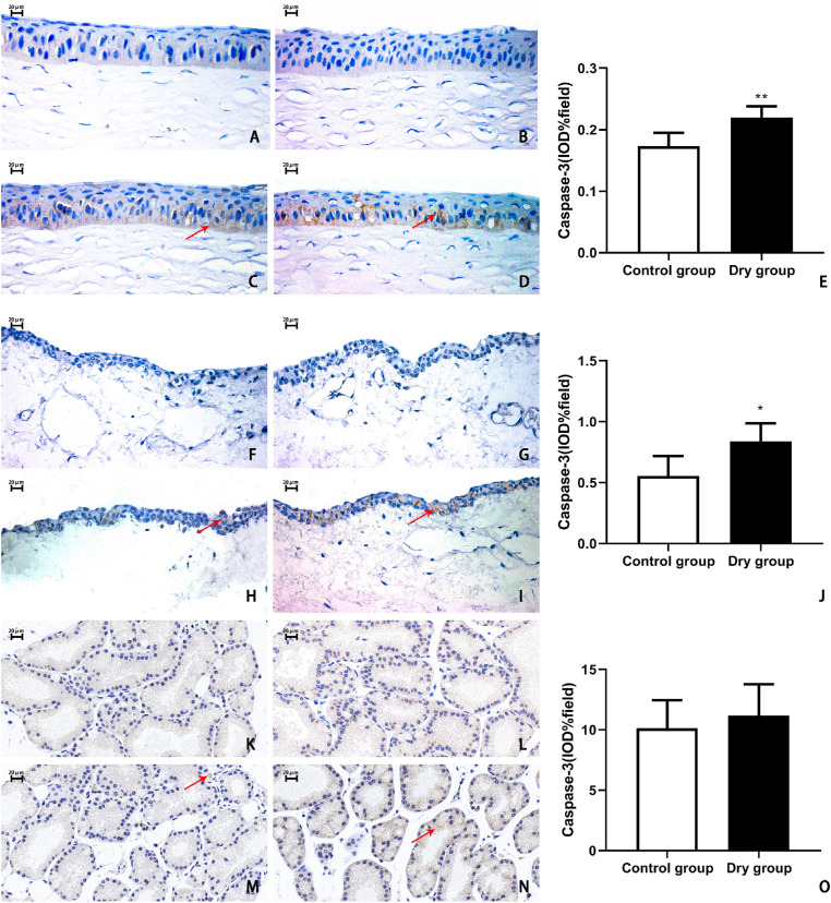
Figure 7.
Representative images and histological analysis of caspase-3 in the corneal epithelium, conjunctival epithelium, and lacrimal gland on day 14. Control experiment showed no positive stained cells in the corneal epithelium (A, B), conjunctival epithelium (F, G), and lacrimal gland (K, I) in the control group and the dry group. On day 14, in the control group, there were little apoptotic cells in the corneal epithelium (C), conjunctival epithelium (H), and lacrimal gland (M). In the dry group, the caspase-3-positive cells in the cornea (D, E) and conjunctiva (I, J) were more significantly increased than in the control group (cornea, **P < 0.01, n = 3 [6 eyes] per group; conjunctiva, *P < 0.05, n = 3 [6 eyes] per group). There were no statistically significant differences in the lacrimal glands (N, O) between these two groups (P > 0.05, n = 3 [6 eyes] each group). Data are shown as mean ± SD (error bars). Magnification, × 400.

