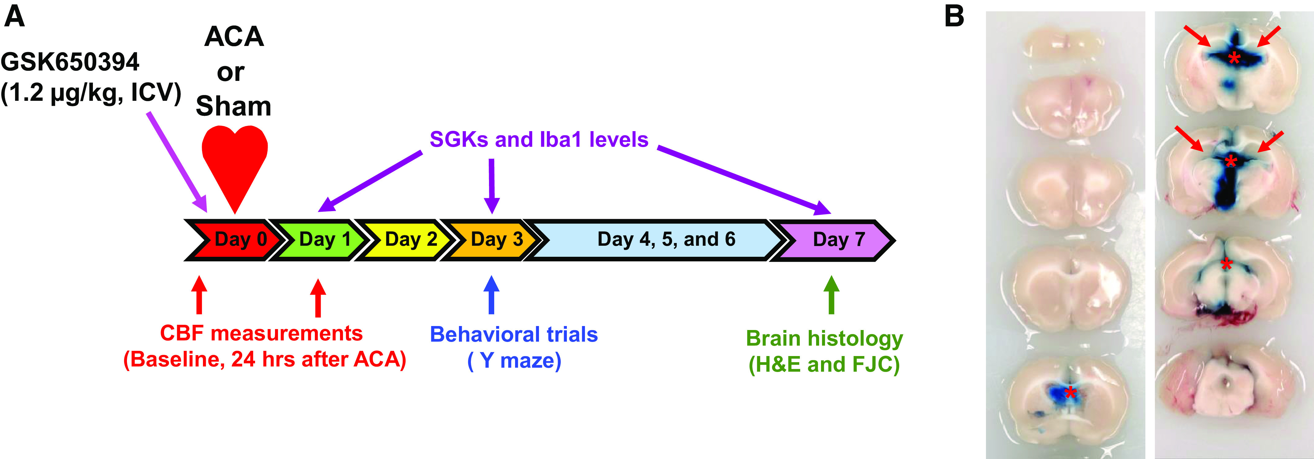
Fig. 2.

Schematic diagram of the experimental design. A: rats received an intracerebroventricular (icv) injection of GSK650394 (1.2 μg/kg) 5 min before asphyxia cardiac arrest (ACA). H&E, hematoxylin and eosin; FJC, Fluoro-Jade C. Cortical cerebral blood flow (CBF) was measured via laser speckle contrast imaging 30 min before and 24 h after ACA to examine the hypoperfusion event. Y maze was implemented 3 days after ACA/sham surgery. Upon completion of behavioral trials, rats were euthanized 7 days after ACA/sham surgery for brain histology. In a separate set of experiments, rats were euthanized 1, 3, and 7 days after ACA/sham for protein and mRNA analyses. SGK1, serum and glucocorticoid-regulated kinase-1. B: representative images of brain sections (2 mm thick) after intracerebroventricular injection of Evans blue. GSK650394 was injected the into third ventricle based on the fact that diffusion of Evans blue (tracer dye) through the ventricular system can be detected in the hippocampus (red arrows). *Evans blue was injected into the third ventricle. The rat was euthanized 5 h after injection.
