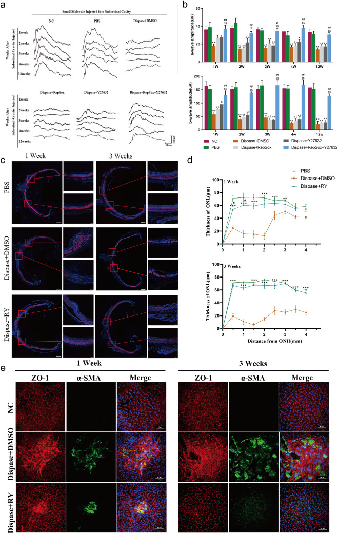
Figure 6.
The combination of RepSox and Y27632 inhibits EMT of RPE cells and maintains visual function in vivo. (a) Representative fERG waves of the normal control (NC), PBS treatment (PBS), dispase plus DMSO treatment (dispase+DMSO), dispase plus RepSox treatment (dispase+RepSox), dispase plus Y27632 treatment (dispase+Y27632), and dispase plus RepSox combined with Y27632 treatment (dispase+RepSox+Y27632) groups at 1, 2, 3, 4, and 12 weeks after injection (PI) were measured by scotopic flash electroretinography (fERG) at a flash intensity of 6.325 × e−2cd*s/m2. (b) Statistical analysis of the amplitudes of fERG a-waves and b-waves at 6.325 × e−2cd*s/m2 in the six groups at one, two, three, four, and 12 weeks PI (n = 8 eyes per group). *P < 0.05, **P < 0.01 versus the PBS group. #P < 0.05, ## P < 0.01 versus the dispase+RepSox group. $P < 0.05, $$P < 0.01 versus the dispase+Y27632 group. (c) Representative images of whole retinal sections (scale bar: 500 µm) that crossed the optic disc were colabeled with the photoreceptor marker recoverin (red) and DAPI (blue). The areas adjacent to the optic disc were collected to show the relative thickness of the outer nuclear layer (ONL) in the three groups at one and three weeks PI. Scale bar: 100 µm. (d) Measurement of the thickness of the ONL in the retinas of S-D rats in the PBS, dispase+DMSO and dispase+RepSox+Y27632 groups at one and three weeks PI (n ≥ 4 eyes per group). ONH: optic nerve head. **P < 0.01, ***P < 0.001 versus the dispase + DMSO group. #P < 0.05 versus the PBS group. (e) RPE-Bruch's membrane choriocapillaris complex images showing the cobblestone-like morphology of RPE cells from the dispase + RepSox + Y27632 group and the disordered appearance of RPE cells from the dispase + DMSO group at one and three weeks PI. RPE cells from the PBS group were used as normal controls. Red: ZO-1; Green: α-SMA; DAPI: nuclei. Scale bar: 50 µm.

