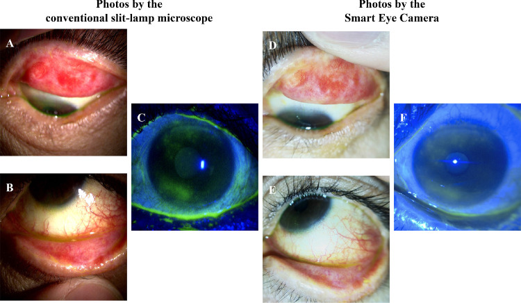
Figure 2.
Comparison of the visual characteristics of dry eye disease between the slit-lamp microscope and the Smart Eye Camera. Clinical photos of the left eye in the same case, which involved a 53-year-old patient with severe ocular graft-versus-host disease with a broad pseudomembrane in the conjunctiva, obvious meibomian gland dysfunction in the lower eyelid, and corneal epitheliopathy. (A), (B), and (C) were recorded using the conventional slit-lamp microscope. (D), (E), and (F) were video recorded using the Smart Eye Camera. A and D show superior tarsal plate with a broad pseudomembrane in the conjunctiva with the diffuse illumination method. B and E show the lower conjunctiva and eyelid with pseudomembranes and meibomian gland dysfunction with the diffuse illumination method. D and F show the corneal epithelial disorder, both with a score of five out of nine points each (upper 2, middle 1, and lower 2) by the blue light illumination method.

