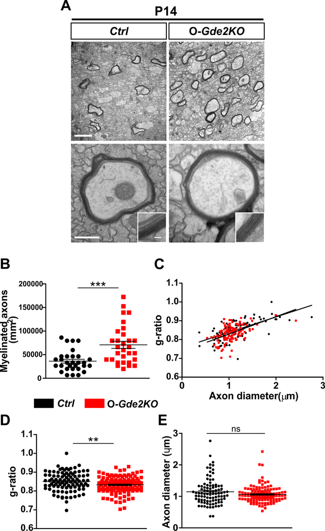
Figure 5. GDE2 ablation in oligodendroglia results in increased myelination.
(A) TEM of P14 CC of Ctrl and O-Gde2KOs. Insets show higher magnification images of the myelin sheath. (B-E) Graphs show an increase in myelinated axons (B, ***p <0.0001; points refer to individual regions of interest [ROI] from 3 Ctrl and 3 O-Gde2KOs), and decrease in g-ratios (C, D **p =0.0056; points refer to individual myelinated axons; 3 Ctrl and 3 O-Gde2KOs) in O-Gde2KO compared with Ctrl but no change in axon diameters (E, ns p = 0.0624; points refer to individual axons, 3 Ctrl and 3 O-Gde2KOs). All graphs: Mean ± sem, two-tailed unpaired Students t-test. Scale bars: (A) 2 μm (top), 500 nm (bottom), inset 50 nm.

