
Fig. 2.
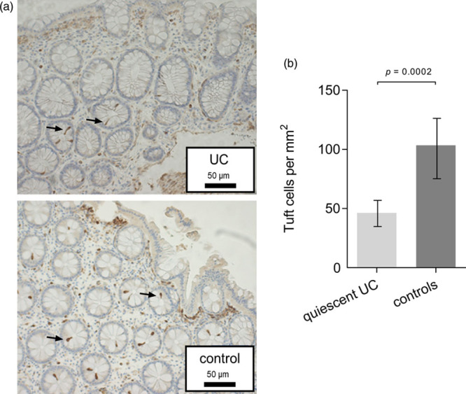
Tuft cells in human sigmoid colon. (a) Examples of immunohistochemical staining of colonic tuft cells with cyclooxygenase-1 specific antibody in a patient with quiescent ulcerative colitis and a control patient. Enlargement × 200. Arrows indicate tuft cells. (b) Colonic mucosal tuft cell numbers per square millimeter in 14 patients with quiescent ulcerative colitis compared to 17 controls. Data presented as mean tuft cells/mm2. The P value is based on Poisson regression with covariate adjustment.