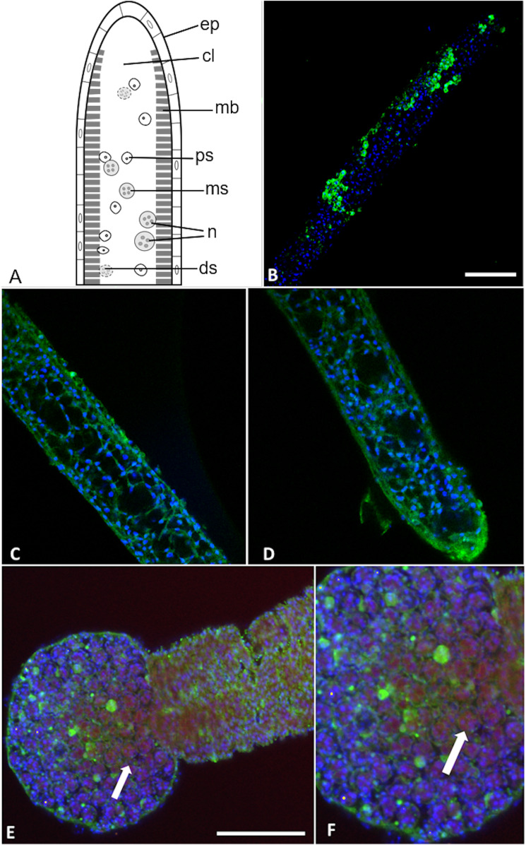
Figure 7. Buddenbrockia plumatellae myxoworms stained with polyclonal antibody raised towards serine peptidase inhibitor toxin.
(A) Schematic of Buddenbrockia plumatellae vermiform stage indicating morphological organisation, ep, epithelium; cl, coelomic cavity; mb, muscle block; ds, developing spores; ms, malacospores; n, nematocyst; ps, presporogonic cells. (B) Serine peptidase inhibitor localised in peripheral cells (green) of presporogonic myxoworm, nuclei stained with DAPI (blue). Scale 60 µm; (C) and (D). Serine peptidase inhibitor (green) localised in cells to the inside of the muscle layer wall, cell nuclei (blue). (E) Spores in distal end of myxoworm with nuclei (blue) and actin (red). Four distinct spherical nematocysts are associated with mature spores. Note, serine peptidase inhibitor (green) localised near body wall surface and not with nematocysts of mature spores (arrow indicating cluster of malacospores). Scale 100 µm; (F). Closer view of (E) showing mature spores with actin (red) in cytoplasm and nuclei (blue), revealing slight staining of serine peptidase inhibitor (green) on surface of spores (arrow).

