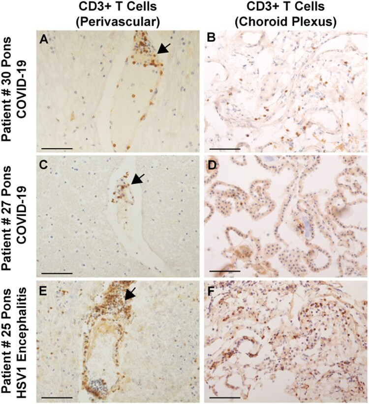
Figure 4.
Immunocytochemical staining for CD3+ T cells in COVID-19 brains. (A and C) Sparse perivascular CD3+ T cells in the pons of COVID-19 Patients 27 and 30. (B and D) Sparse CD3+ T cells in the choroid plexus from the lateral ventricle of Patients 27 and 30. (E and F) CD3+ T cell infiltrates around a pontine vessel (E) and in the choroid plexus (F) of Patient 25 with HSV-1 encephalitis; arrows indicate CD3+ T cell infiltrates. All sections are counterstained with haematoxylin. Scale bars = 200 µm.

