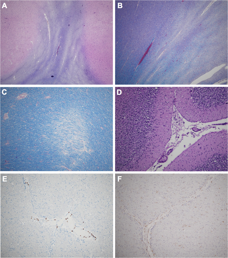
Figure 1.
Histologic features of Case 1. LFB/PAS stains demonstrating serpiginous myelin pallor in the temporal (A) and peri-hippocampal (B) white matter. Higher-power LFB/PAS demonstrating perivascular myelin loss (C). Loss of Purkinje cells and Bergmann gliosis (D). There are multifocal regions of perivascular CD3+ T-lymphocytes (E) and perivascular and parenchymal CD68+ histiocytes/activated microglial cells (F).

