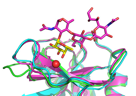
Fig. 8.

Comparison of structure of E-selectin (low-affinity conformation) soaked with sLeX published by Somers et al. (green), structure of E-selectin in high-affinity conformation co-crystalized with sLeX published by Preston et al. (cyan) and prepared high-affinity conformation used for docking (magenta) (Somers et al. 2000; Woelke et al. 2013; Preston et al. 2016). The fucose residue is shown in yellow and the Ca2+ cation as a red sphere. This figure is available in black and white in print and in color at Glycobiology online.
