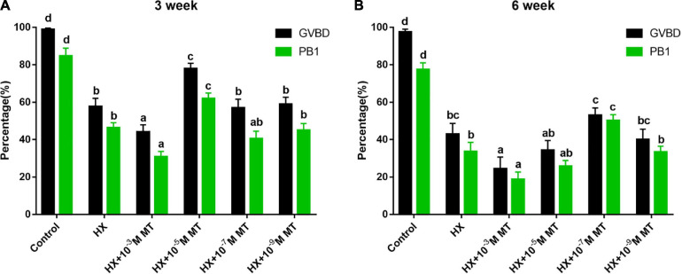
FIGURE 1.
The effect of melatonin on the meiosis of mouse oocytes in the presence of HX. GVBD and PB1 during oocyte maturation. (A) Three-week-old. Control (n = 153), HX (n = 153), HX + 10–3 M, MT (n = 159), HX + 10–5 M, MT (n = 154), HX + 10–7 M MT (n = 158), HX + 10–9 M MT (n = 161). (B) Six-week-old mice. Control (n = 152), HX (n = 148), HX + 10–3 M MT (n = 149), HX + 10–5 M MT (n = 158), HX + 10–7 M MT (n = 152), HX + 10–9 M MT (n = 153). HX, 4 mM hypoxanthine; MT, melatonin. Data represents the mean with SEM. Different superscript letters (a–d) in each column present significant differences. (P < 0.05) determined by one-way ANOVA followed by Tukey post hoc comparisons.

