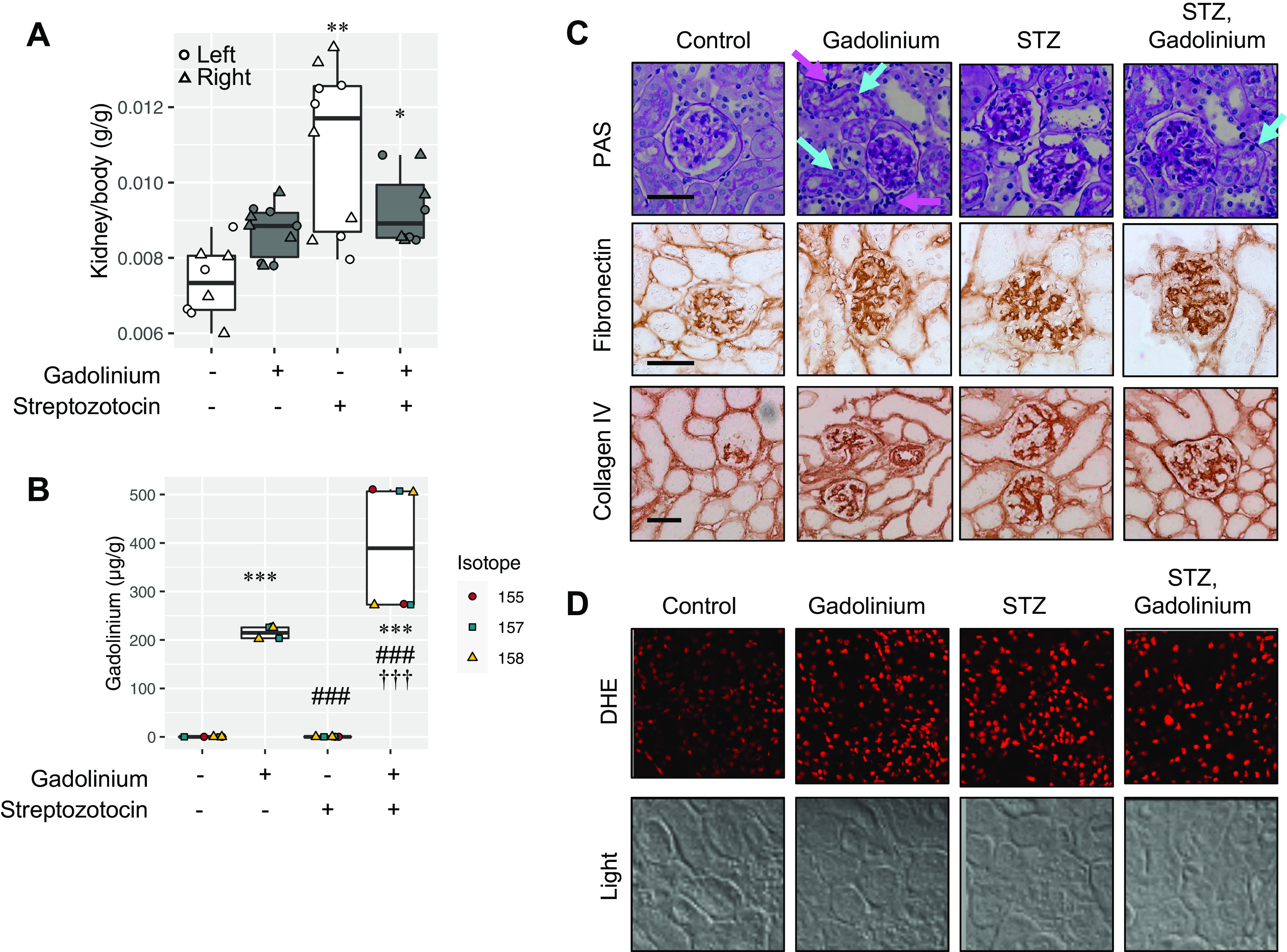
Figure 3.

Superimposition of streptozotocin (STZ)-induced experimental diabetes and gadolinium-based contrast agent treatment in the kidney. A: kidney weights for untreated (n = 8), gadolinium (n = 10), STZ (n = 10), and the combination of STZ and gadolinium (n = 8). Left kidney (circle), right kidney (triangle). B: experimentally induced diabetes increased gadolinium retention in the kidney. Analysis of variance and Tukey honestly significant difference by post hoc test (*P < 0.05, **P < 0.01, and ***P < 0.001 from the first lane, ###P < 0.001 from the second lane, and †††P < 0.001 from third lane). C: impact of gadolinium-based contrast agent treatment and experimental diabetes in the renal cortex. Top, gadolinium-based contrast agent treatment-induced tubular damage (cyan arrows) and increased interstitial cells with dense nuclei (magenta arrows) in addition to increased glomerular mesangial matrix. The combination of experimental diabetes and contrast treatment was characterized by mesangial expansion and acute tubular damage (with heterogenous proximal tubular nuclei, cyan arrows). Periodic acid-Schiff (PAS) staining is shown. Middle, fibronectin expression. Immunohistochemistry was performed. Bottom, collagen type IV staining. Immunohistochemistry was performed. Calibration bars = 0.05 mm. D: gadolinium-based contrast agent treatment and experimental diabetes increased renal cortical reactive oxygen species generation. DHE, dihydroethidium. Overall magnification: ×200.
