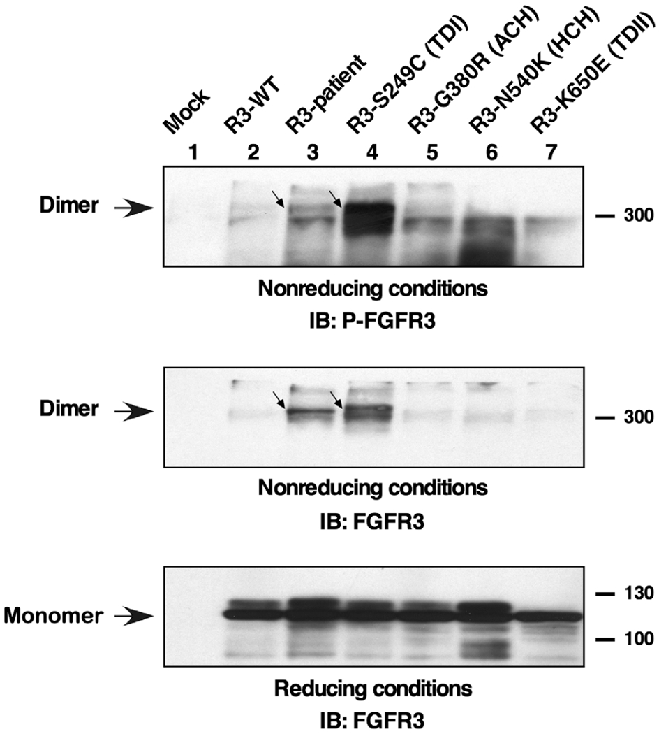
FIGURE 4.

Tyrosine phosphorylation of FGFR3 dimers. Lysates from HEK293 cells expressing FGFR3 derivatives were resolved by 4–12% gradient SDS-PAGE under nonreducing conditions and immunoblotted with antisera specific for phosphorylation of the activation loop tyrosine residues of FGFR3. Phosphorylated dimers are indicated (top panel). The same membrane was reprobed for total FGFR3 (middle panel). Duplicate samples were analyzed under reducing conditions and immunoblotted for FGFR3 showing the total of monomer receptors (bottom panel)
