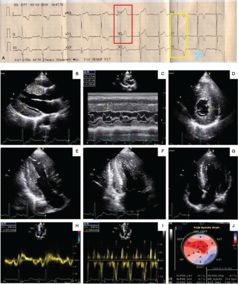
Figure 1.

Surface electrocardiogram of the patient showing evidence of left ventricular hypertrophy (red box and yellow box) with strain pattern (arrow) (A). Trans-thoracic echocardiography showing evidence of asymmetric LVH (B) with inter-ventricular septal diameter of 2.4 cm (yellow arrow) in diastole (C) along with speckled pattern in the myocardium (D, E) without any evidence of left ventricular outflow tract gradient or systolic anterior motion of anterior mitral valve leaflet (F, G). Tissue Doppler study revealed grade 2 left ventricular diastolic dysfunction (H, I). Strain echocardiography using speckled tracking revealed marked diminution of global longitudinal strain with apical sparing (J). LVH = left ventricular hypertrophy.
