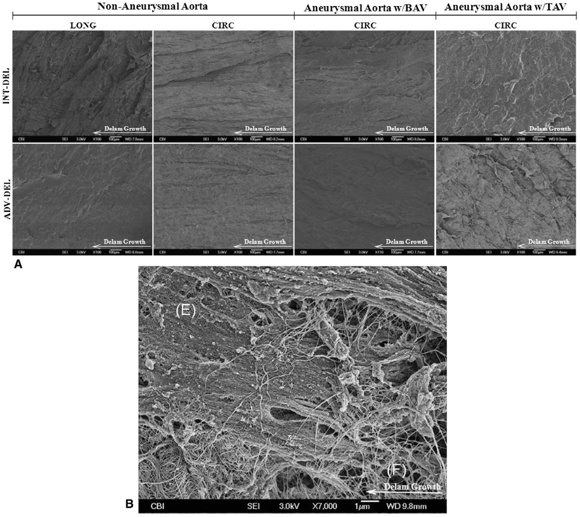
FIGURE 4.

A, Representative scanning electron microscope (SEM) images of fracture surfaces of nonaneurysmal and ascending thoracic aortic aneurysm (ATAA) with bicuspid aortic valve (BAV) and tricuspid aortic valve (TAV) in longitudinal (LONG) and circumferential (CIRC) direction. B, High magnification image of CIRC TAV ATAA for intimal surface and delaminated plane (INT-DEL) half showing bundles of broken elastin fibers (F) existing between elastic sheets (E). Fibers act as bridge between halves in fracture modality called “fiber bridging” in delamination testing.
