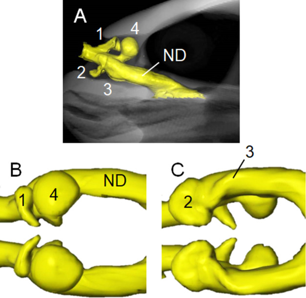
Fig 2. Three-dimensional reconstructions based on computed tomography images of nasal cavity of hawksbill sea turtle.
(A) Left lateral view of internal nasal cavity (yellow) at anterior region of skull. Dorsal (B) and ventral (C) views of nasal cavity. Anterodorsal (1), posterodorsal (4) and anteroventral (2) diverticula, and small posteroventral salience formed by fossa on wall (3) in cavum nasi proprium anterior to nasopharyngeal duct (ND). See also S1 Video.

