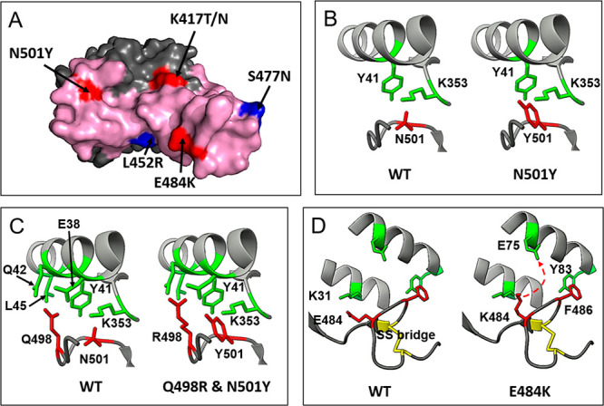
Figure 2.

Selected mutations in RBD. (A) Location of each mutated amino acid with respect to the RBM surface (pink). Residues belonging to the ACE2 epitope are highlighted in red, and those without contact with ACE2 are colored in blue. (B) and (D) New interactions associated with N501Y and E484K mutations, respectively. (C) Potential epistatic mutation Q498R associated with N501Y.
