Figure 1.
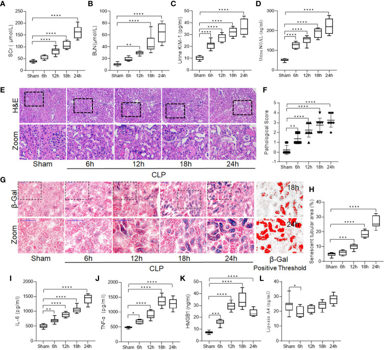
Increased renal damage was accompanied by enhanced tubular cell senescence and inflammation in sepsis induced-AKI. Male Sprague-Dawley rats underwent CLP were sacrificed at the time point of 6-h, 12-h, 18-h, 24-h after surgery. Renal function alternation, renal necrosis and renal tubular epithelial cells senescence were detected with different methods in kidneys. (A–D) levels of SCr, BUN, urinary KIM-1 and NGAL at different surgery time points after CLP. (E) Renal damage of rats at different surgery time points after CLP (H&E; scale bar 100 μm). (F) Kidney histopathology evaluation scores at different surgery time points. (G) Representative images of cellular senescence in kidneys after CLP (β-Gal staining; scale bar 100 μm); Red: β-Gal positive threshold. (H) Senescent tubular areas at different surgery time points after CLP. (I–K) levels of IL-6, TNF-α and HMGB1 at different surgery time points after CLP. (L) levels of lipoxin A4 at different surgery time points after CLP. Data are presented as mean ± SE (n = 8). *p < 0.05; **p < 0.005; ***p < 0.001; ****p < 0.0001; CLP, cecal ligation and puncture; AKI, acute kidney injury; H&E, hematoxylin–eosin staining; SCr, serum creatinine; BUN, blood urea nitrogen; KIM-1, kidney injury molecule-1; NGAL, neutrophil gelatinase-associated lipocalin; IL-6, interlecukin-6; IL-8, interlecukin-8; TNF-α, tumor necrosis factor-alpha; LXA4, lipoxin A4.

