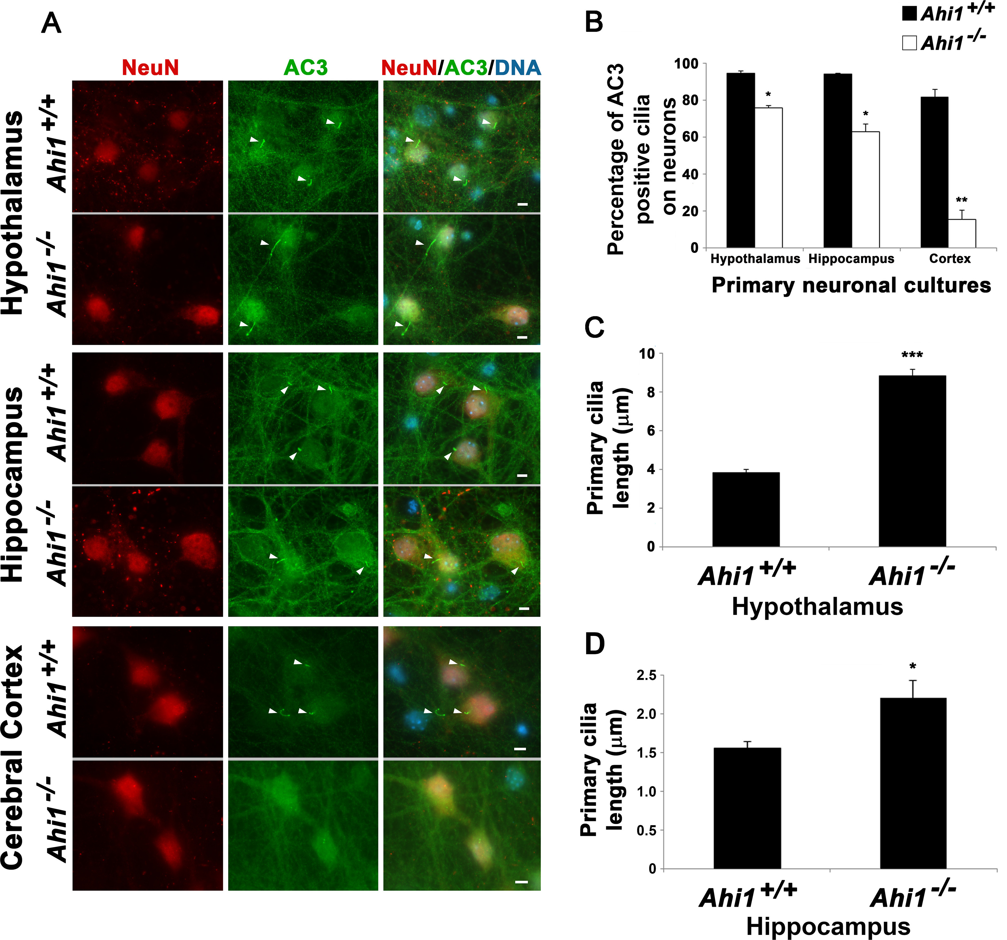
Figure 3.

Loss of Ahi1 results in reduced AC3-positive cilia number and cilia elongation in neurons. A, Primary cilia on Ahi1+/+ and Ahi1–/– neurons were identified by performing co-immunolabeling of AC3 (ciliary marker) and NeuN (neuronal marker). DNA was visualized with Hoechst 33258 (blue). Analysis was performed in neurons isolated from three different brain areas: hypothalamus, hippocampus, and cerebral cortex. White arrowheads point to primary cilia. Scale bar: 5 µm. B, Percentage of neurons with AC3-positive cilia in the three different brain areas; n ≥ 100 neuronal cells from each embryo (n = 3 per tissue/genotype). Error bars represent SEM. Significance was determined by χ2 tests (*p < 0.05, **p < 0.005). C, D, Cilia length analysis from Ahi1+/+ and Ahi1–/– hypothalamic (C) and hippocampal (D) neurons. Primary cilia lengths were measured using AxioVison software, n ≥ 100 (C) and n ≥ 20 (D) neuronal cells/group. Error bars represent SEM. Significance was determined by unpaired two-tailed t tests (*p < 0.03, ***p < 0.0005).
