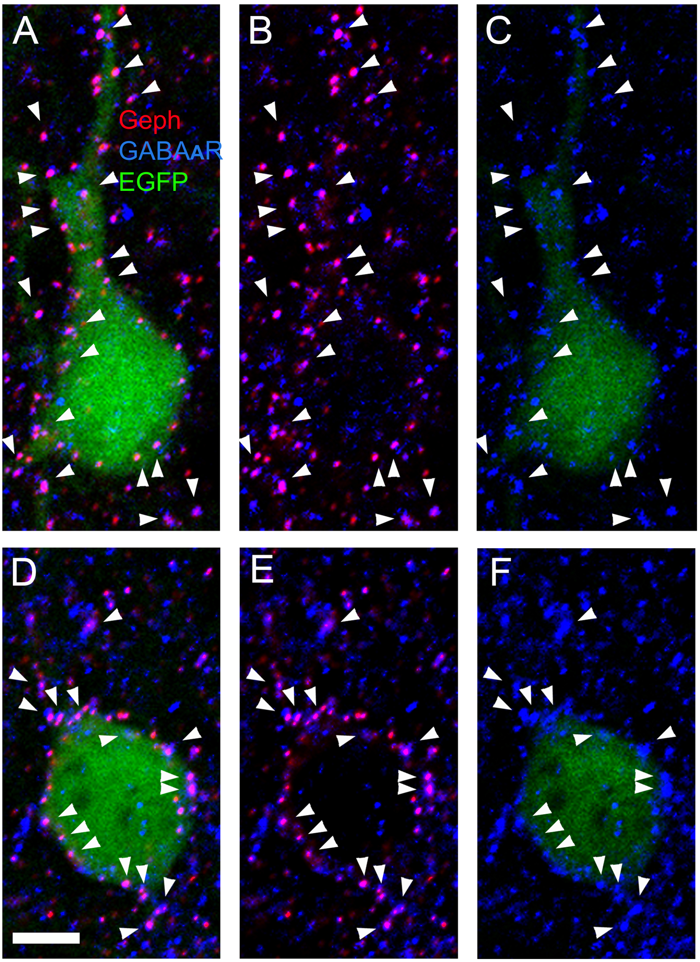
Figure 6. In CB2SH3− overexpressing neurons, GABAARs co-localize with gephyrin superclusters.

(A-C and D-F) Triple-label fluorescence with anti-Geph (red), anti-γ2 GABAAR subunit (blue) and EGFP (green) of two transfected cortical neurons at P45. A and D show red, green and blue overlays. B and E show red and blue overlays. C and F show green and blue overlays. Arrowheads point to co-localizing Geph and GABAAR clusters. Scale bar = 5 μm.
