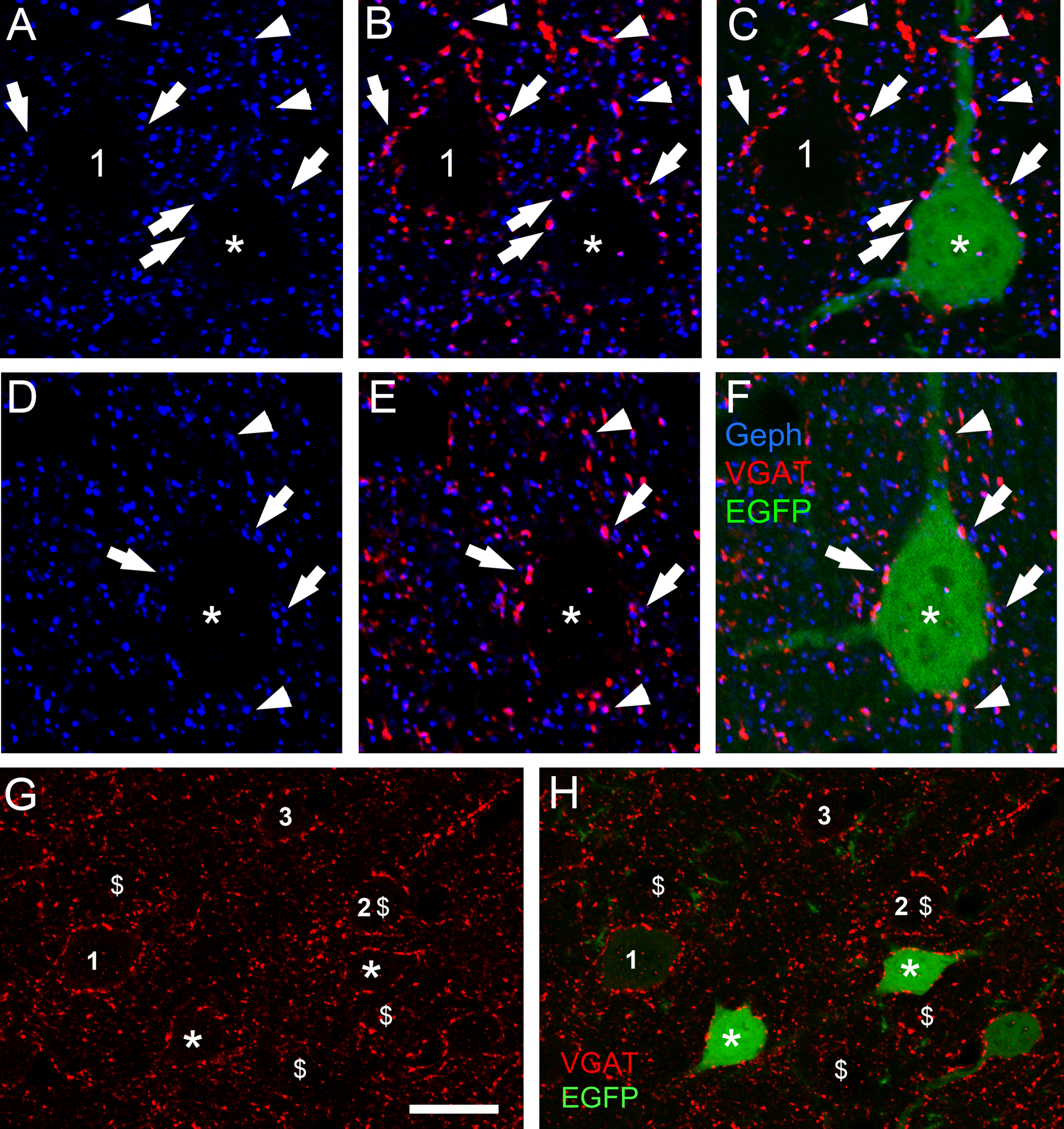
Figure 9. In CB2SH3+ overexpressing neurons, gephyrin clusters are associated with presynaptic vGAT puncta. There is no effect on the extent or fluorescence intensity of the vGAT puncta contacting the transfected neuron.

(A-C and D-F) Triple-label fluorescence with anti-Geph (blue), anti-vGAT (red) and EGFP (green) at P42 cortical neurons. Arrows and arrowheads show some examples of apposition of Geph clusters and vGAT in somas (arrows) and dendrites (arrowheads). C and F show the RGB overlays. For comparative purposes A-C shows a transfected neuron (green, asterisk) and a non-transfected sister neuron (number 1). (G and H) Double-label fluorescence. The extent and fluorescence intensity of vGAT puncta (red) contacting CB2SH3+ transfected neurons (green, asterisks) and non-transfected neurons (numbers 1–3) are similar. Note that the transfected neurons (green) in G and H are the same as in Fig 7A and B. For comparative purposes $ symbols were added corresponding to the cells labelled with the $ symbol in Fig 7A and B. Scale bar = 10 μm in A-F and 17 μm in G and H.
