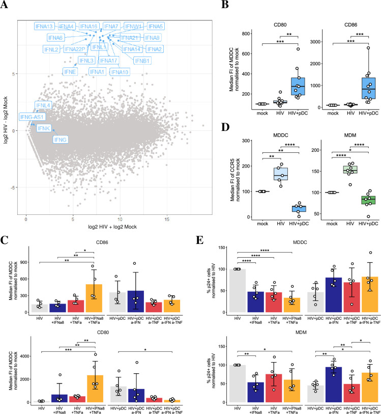
Fig 2. Mechanisms of reduced infectivity in MDDCs and MDMs.
(A) RNAseq data showing IFN-subtype gene induction in pDCs at 18 hours post HIV exposure versus mock. (B-C) The median FI of maturation markers, CD80 (n = 9) and CD86 (n = 10) in MDDCs that were either mock, HIV infected MDDCs in the presence and absence of pDCs, and in HIV infected MDDCs treated with exogenous rIFNα8 and/or rTNFα, HIV infected MDDCs treated with antibodies to blocking IFN and/or TNF signaling in pDC cocultures (n = 4 for CD80, n = 5 for CD86). Data is shown as normalized to 100% in mock infected. (D) pDC decreased CCR5 median FI in HIV-infected MDDCs (n = 5) and MDMs (n = 7). Data is shown as normalized to 100% in mock infected. (E) Changes in p24 expression at 5 dpi in MDDCs (n = 5) (top panel) and MDMs (n = 5) (bottom panel) upon addition of exogenous rIFNα8 and/or rTNFα, or upon blocking IFN and/or TNF signaling in pDC cocultures. Data was normalized to 100% in HIV infected cells in the absence of pDCs. For all data, *p < 0.05, **p < 0.01, ***p < 0.001, ****p < 0.0001 by repeated measures ANOVA with Tukey post-hoc test.

