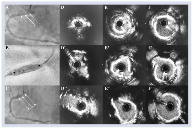
Figure 1.
A. Subtotal occlusion of the ostial right coronary artery (RCA) with tortuous uptake from the aorta; B. Angiographic appearance of the Shockwave intravascular lithotripsy (IVL) balloon; C. Final angiographic result after stent implantation; D–F. Intravascular ultrasound (IVUS) before IVL revealing three-to-four-quadrant (270° to 360°) calcification of the proximal RCA along with severe ostial stenosis; D’–F’. IVUS after IVL demonstrating successful fracture of the calcified lesion within proximal RCA; D”–F”. Final IVUS showing optimal stent expansion.

