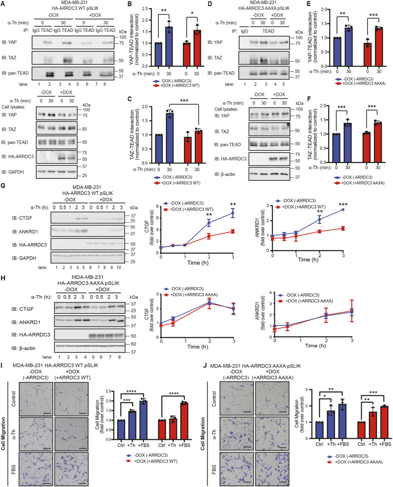
Fig. 7.
ARRDC3 re-expression inhibits TAZ–TEAD binding and attenuates downstream Hippo signaling and thrombin-induced migration, dependent on the PPXY motifs of ARRDC3. (A–H) MDA-MB-231 wild-type (WT; A–C,G) and AAXA mutant (D–F,H) HA–ARRDC3 pSLIK cells were treated with doxycycline (DOX) to induce ARRDC3 expression and stimulated with 10 nM α-thrombin (α-Th) for indicated times. (A–F) Cells were lysed and immunoprecipitated (IP) with anti-TEAD antibody or anti-IgG control. IP samples and cell lysates were immunoblotted (IB) with antibodies against the indicated proteins. Results are quantified, and co-association of YAP–TEAD (B,E) and TAZ–TEAD (C,F) is represented as fold over −DOX 0 min control. Data are mean±s.d., n=3. Statistical significance determined using two-way ANOVA with Tukey′s post hoc test. (G,H) Results are the fold-change in CTGF and ANKRD1 expression relative to 0 min −DOX control. Data are mean±s.d., n=3. Statistical significance was determined by unpaired t-test at each time point. (I,J) Migration assay with MDA-MB-231 wild-type (I) and AAXA mutant (J) HA–ARRDC3 pSLIK cells treated with doxycycline to induce ARRDC3 expression and incubated with or without 100 pM α-thrombin or 0.5% FBS. Images shown are representative of three independent experiments. Scale bars: 20 μm. Results are the fold change over untreated control cells. Data are mean±s.d., n=3. Statistical significance was determined by one-way ANOVA with Tukey′s post hoc test. *P<0.05; **P<0.01; ***P<0.001; ****P<0.0001.

