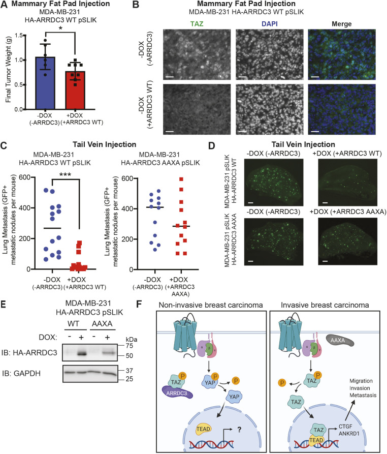
Fig. 8.
ARRDC3 re-expression blocks in vivo breast cancer growth and metastasis, dependent on the PPXY motifs of ARRDC3. (A,B) MDA-MB-231 wild-type (WT) HA–ARRDC3 pSLIK cells were injected in the mammary fat pad of NSG mice fed with or without doxycycline (DOX). (A) Final tumor weight 6 weeks post-implantation. Statistical significance determined by unpaired t-test (mean±s.d.; n=6 mice in −DOX group, n=8 mice in +DOX group). (B) Representative images of immunohistochemistry of mammary fat pad tumors stained for TAZ (green) and DAPI for nuclei (blue). Scale bars: 25 µm. (C,D) GFP-labeled MDA-MB-231 wild-type or AAXA mutant HA–ARRDC3 pSLIK cells were injected into the tail vein of NSG mice. (C) Quantification of GFP-positive metastatic nodules in the lungs of the mice collected 2 weeks after injection. Line indicates the median. Statistical significance determined by unpaired t-test with Welch's correction (wild type, n=14 mice per group; AAXA, n=12 mice per group). (D) Representative fluorescence images of GFP-positive metastatic lesions in the lungs of mice. GFP signal indicates tumor cell extravasation, seeding, growth and colonization in the lung. Scale bars: 1 mm. (E) Verification of HA–ARRDC3 wild-type or HA–ARRDC3 AAXA re-expression in MDA-MB-231 pSLIK cells collected prior to tail-vein injection. Lysates immunoblotted (IB) for HA–ARRDC3 and GAPDH expression. (F) ARRDC3 is highly expressed in normal mammary epithelial cells or luminal non-invasive breast carcinoma cells, and co-associates with TAZ, leading to its cytoplasmic retention and attenuated GPCR-mediated Hippo pathway signaling. However, when ARRDC3 is absent, e.g. in invasive basal-like breast carcinoma, or is lacking PPXY motifs and thus functionally inactive, GPCR signaling activates TAZ through TAZ dephosphorylation, promoting its nuclear localization, binding to the TEAD family of transcriptional co-activators, and inducing CTGF and ANKRD1 expression, leading to cell migration, invasion and metastasis. P, phosphorylation. Model created with BioRender.com. *P<0.01; ***P<0.001.

