Fig. 2.
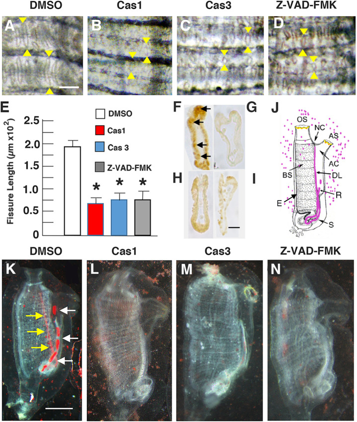
Apoptosis is required for branchial sac homeostasis and function. (A–D) Branchial sac fissures after 6-day treatment with (A) DMSO (control) (B) caspase 1 inhibitor, (C), caspase 3 inhibitor, or (D) pan-caspase inhibitor Z-VAD-FMK. Arrowheads: distal (top) and proximal (bottom) ends of the fissures. Scale bar: 15 µm; magnification is the same in A–D. (E) Bar graphs showing the mean lengths of branchial sac fissures in caspase inhibitor and DMSO (control) treated animals. N=12 for each bar. Error bars: s.e.m. Asterisks indicate significant difference at P=0.004 between the control and caspase inhibitor treated animals. Statistics by one-way ANOVA and post-hoc Tukey with Bonferroni correction. (F–I) Sections of TUNEL assayed pharyngeal fissures of (F) DMSO (control), (G) caspase 1 inhibitor-, (H) caspase 3 inhibitor, or (I) pan-caspase inhibitor Z-VAD-FMK treated animals. Arrows in F: TUNEL labeled cells. Scale bar: 5 µM; magnification is the same in all frames. (J) Diagram illustrating the carmine particle assay for branchial fissure function. Carmine particles shown by magenta dots and colored organs in the body. OS, oral siphon; AS, atrial siphon; NC, neural complex; BS, branchial sac; DL, dorsal lamina; S, stomach; R, rectum; AC, atrial cavity. (K–N) Carmine particle assay of animals treated with (K) DMSO, (L) caspase 1 inhibitor, (M) caspase 3 inhibitor, or (N) pan-caspase inhibitor Z-VAD-FMK. Scale bar: 110 µm; magnification is the same in K–N. Arrows in K show carmine particles concentrated in the dorsal lamina (yellow arrows) and rectum (white arrows). Each experiment was replicated at least three times.

