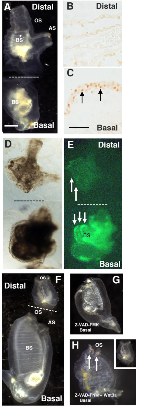
Fig. 6.

The roles of apoptosis-dependent regeneration and stem cell activation in mid-body amputation. (A) Bisected animal immediately after mid-body amputation showing distal and contracted basal fragments. Scale bar in A: 100 µm. Magnifications are the same in A and D–H. (B,C) Sections of the distal (B) and basal (C) fragments 12 h after mid-body amputation showing TUNEL labeled cells (arrows) in the basal but not the distal fragment. Scale bar in C: 10 µm. Magnifications are the same in B and C. (D,E) Bright field (D) and fluorescence (E) images of basal and distal fragments subjected to EdU for 2 days after bisection showing progenitor cell labeling in the branchial sac stem cells of the basal (downward arrows) but not the distal fragment (upward arrows). (F) Bisected control animal after 6 days PA showing the regenerating basal fragment and non-regenerating distal fragment. (G) A basal fragment treated with pan-caspase inhibitor Z-VAD-FMK immediately after mid-body amputation showing the absence of regeneration at 6 days PA. (H) A basal fragment treated with pan-caspase inhibitor Z-VAD-FMK and Wnt3a immediately after mid-body amputation showing rescue of regeneration in the basal fragment (arrows) but not the distal fragment (inset) at 6 days PA. OS, oral siphon; AS, atrial siphon; BS, branchial sac. Dashed lines in A, D–F indicate the bisection plane. Each experiment was replicated at least three times.
