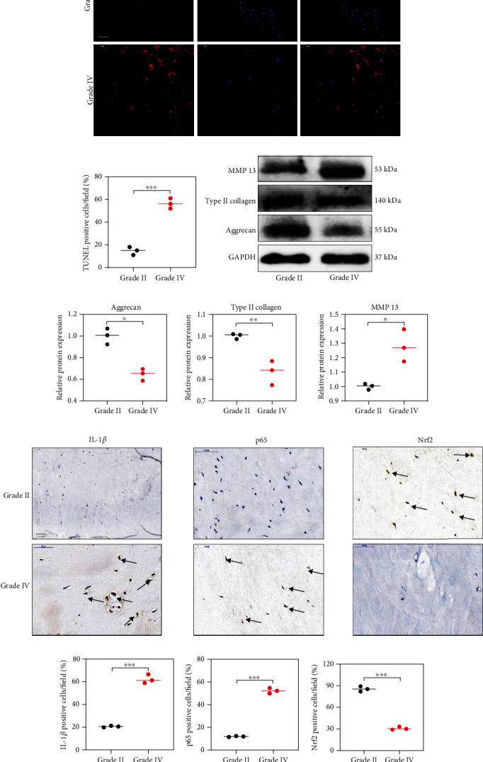
Figure 1.

The protein expression of Nrf2 and p65 in human NP tissues. (a) The apoptotic NP cells (red) in intervertebral disc between Grade II and Grade IV were visualized via TUNEL staining and the nuclei were stained with DAPI. Scar bar = 300 μm. (b) Three randomized versions were selected, and TUNEL staining-positive cells were quantified via the amount of red fluorescence. (c, d) Protein bands and quantification of expression levels of Aggrecan, Type II collagen, and MMP 13. Scar bar = 100 μm. (e) Immunohistochemical results were used to examine the protein levels of IL-1β, p65, and Nrf2 in the human NP tissue with Grade II and Grade IV. (f) Three versions were randomly selected, and the stained cells were quantified separately. ∗p < 0.05, ∗∗p < 0.01, ∗∗∗p < 0.001, ∗∗∗∗p < 0.0001.
