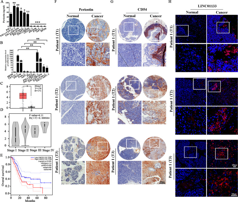
Fig. 3. Increased LINC01133 expression positively correlates with Periostin and exosomal CD54 expression, as well as with poor PDAC patient survival.
A Periostin expression in PSCs was enhanced when PSCs were co-cultured with PCCs. The experiment was repeated three times, and the significance was analyzed by a Student’s t test. Data are shown as mean ± SD (*P < 0.05, **P < 0.01 and ***P < 0.001 vs. PSCs). B LINC01133 expression in PCCs was also enhanced when PCCs were co-cultured with PSCs. The experiment was repeated three times, and the significance was analyzed using a Student’s t test. Data are shown as mean ± SD (**P < 0.01, PCCs vs. PSCs + PCCs.). C TCGA database analysis suggested that LINC01133 had significantly higher expression in PDAC tissues (P < 0.05). D TCGA database analysis also suggested that LINC01133 was significantly positively corelated with the TNM stage of PDAC patients (P < 0.001). E High expression of LINC01133 was significantly positively correlated with poor overall survival of PDAC patients, as suggested by TCGA database analysis (P = 0.028). F, G Immunohistochemical staining of 80 paired pancreatic cancer and matched normal tissues with anti-periostin and anti-exosomal CD54 antibodies and the representative patient samples of clinical stages T1, T2, and T3 are shown. H Representative images of LINC01133 expression (red, detected by RNA-FISH) in the same tumor tissue and adjacent non-tumor tissues.

