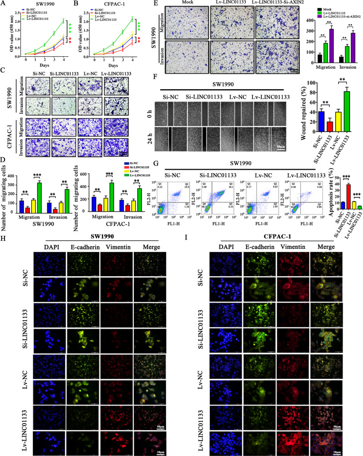
Fig. 4. LINC01133 promotes pancreatic cancer cell proliferation, migration, invasion, and EMT, and decreases pancreatic cancer cell apoptosis.
A, B LINC01133 knockdown decreased the proliferation rates of SW1990 and CFPAC-1 cells. In contrast, increased LINC01133 expression accelerated the proliferation rates of SW1990 and CFPAC-1 cells. OD values at 450 nm were measured by CCK-8 assay at 0, 1, 2, 3, and 4 days. The experiment was repeated three times, and the significance was analyzed by a Student’s t test. Data are shown as mean ± SD (**P < 0.01 and ***P < 0.001 vs. NC). C, D LINC01133 knockdown inhibited the migration and invasion rates of SW1990 and CFPAC-1 cells, whereas enhanced LINC01133 expression exerted the opposite effect. Cells were stained with crystal violet, and representative photographs of migratory or invading cells on the membrane coated with or without Matrigel (×50 magnification; Zeiss) are shown. The experiment was repeated three times, and the significance was analyzed by a Student’s t test. Data are shown as mean ± SD (**P < 0.01 and *** P < 0.001 vs. NC). E The migration and invasion ability were enhanced in AXIN2 silenced Lv-LINC01133 SW1990 cells. The experiment was repeated three times, and the significance was analyzed by a Student’s t test. Data are shown as mean ± SD (**P < 0.01 vs. Mock). F Migration activity was measured by wound-healing assay and the migration distance was measured from five random fields captured at each indicated time point. The experiment was repeated three times, and wound repair percentage of each cell line is shown in bar charts as mean ± SD (**P < 0.01 vs. NC). G Apoptosis was evaluated in SW1990 transfected with Si-NC, Si-LINC01133, Lv-NC and Lv-LINC01133 by Annexin-V-PI staining and flow cytometry. The apoptosis experiment was repeated three times, and the representative histograms are shown. The apoptosis rates are shown as mean ± SD (***P < 0.001 vs. NC). H–I The EMT effect was further validated by immunofluorescence analysis in SW1990 and CFPAC-1 cells.

