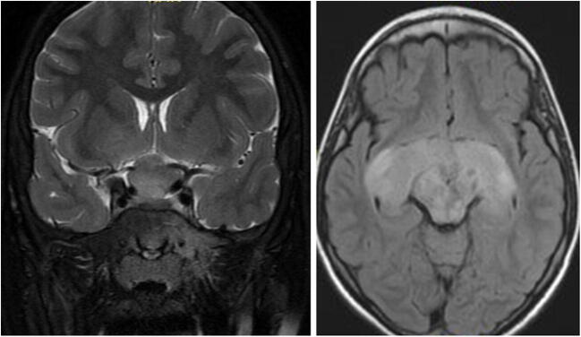
Fig. 4.
Optic pathway glioma (OPG) in neurofibromatosis type I (NF1): coronal T2-weighted MRI and axial FLAIR MRI of a 9-year-old boy with NF1. MRI shows a typical OPG, involving the optic nerves, chiasm, and optic tracts. Typical NF changes are seen in the mid-brain. The child underwent treatment with vincristine and carboplatin, followed by vinblastine. Over the years, the tumor reduced in size; however, vision continued to deteriorate

