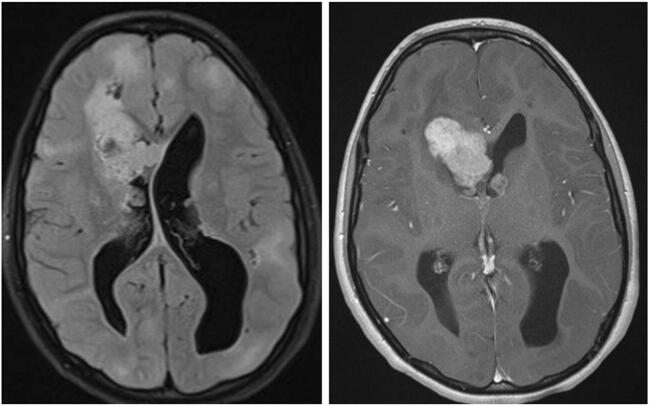
Fig. 5.
Subependymal giant cell astrocytoma (SEGA): axial FLAIR and T1-contrast-enhanced MRI of a 12-year-old girl with known tuberous sclerosis and epilepsy, presenting with new-onset symptoms of increased intracranial pressure. MRI shows multiple cortical tubers, and bilateral SEGA (prominent on the right side) with obstructive hydrocephalus. The child underwent transcortical resection of the tumor and a septostomy. No progression of the left tumor or recurrence of the right tumor is evident over a 6-year follow-up

