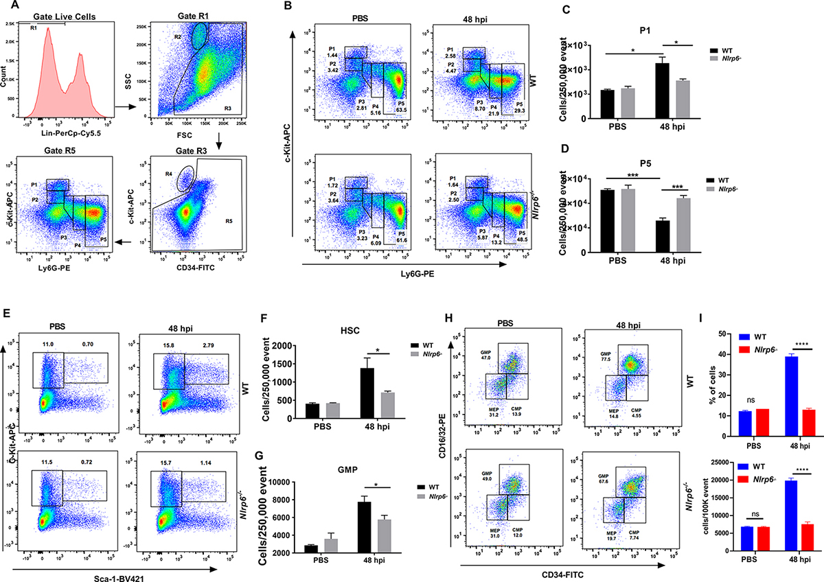
Figure 4. Role of NLRP6 in emergency granulopoiesis and neutrophil release during Kp-induced lung infection.

(A–H) WT and Nlrp6−/− mice were infected with Kp (103 CFU/mouse) and lungs were harvested at 48 h post-infection. (A) Flow cytometric analysis of granulopoiesis. First, BM cells that are lineage positive for B, T and erythroid cells were excluded as they have lost the potential to give rise to granulocytes. The remainder, gate R5, was then plotted against expression of c-Kit and Ly-6G. Populations R2 and R4 represent eosinophilic and megakaryocyte-erythroid progenitors, respectively. FACS dot plot (B), percentage of subpopulation #1 (C), and subpopulation #5 (D) within the granulopoietic compartment are presented. FACS analysis plot (E) and number (F) of hematopoietic stem cells (HSC)(c-Kit+Sca-1+Lin−) and FACS dot plot (G) number (H) of granulocyte-monocyte progenitor (GMP) cells (within c-Kit+Sca-1−Lin−) at 48 h post-infection. (n= 4–6 mice/infection group, n=3 mice/control group). (I) FACS analysis of blood neutrophils at 48 h post-infection with Kp (n=6 mice/group). The % and number of cells were determined after PBS instillation or infection with Kp. Statistical significance was determined by unpaired t-test (C, D, F, G). *p<0.05; ***, p<0.001. CMP: Common myeloid progenitors, MEP: Megakaryocyte-erythroid progenitors.
