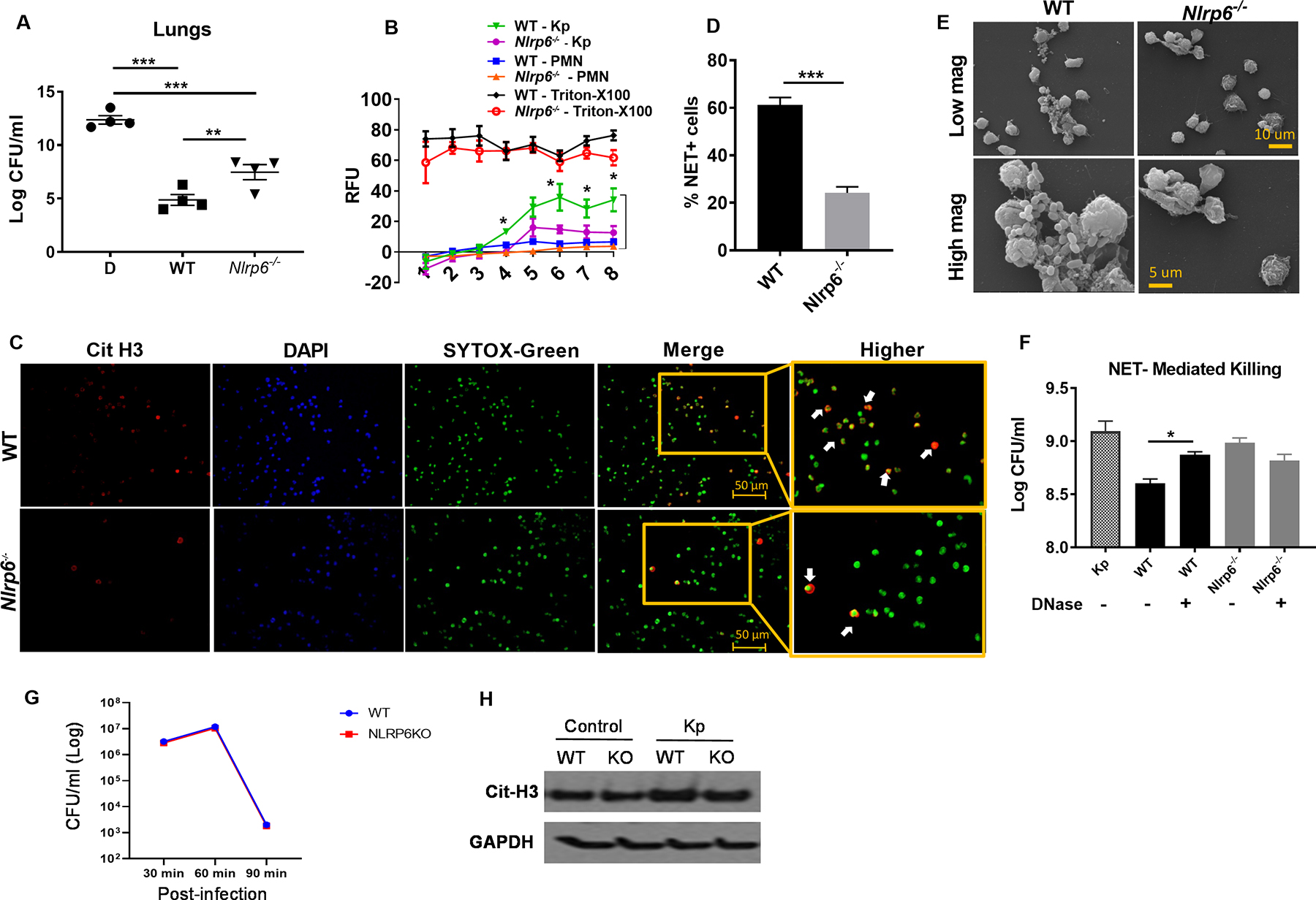
Figure 5. The effect of NLRP6 on NETosis and NET-mediated bacterial killing.

(A) In Nlrp6−/− mice, neutrophils were depleted by administration of anti-Ly6G mAb intraperitoneally and replenished with freshly isolated BMDNs (2 ×10⁶ cells/mouse) from WT or Nlrp6−/− mice i.t. 30 min prior to Kp infection. At 48 h post-infection, bacterial burden was assessed in lungs (n=4/group). (B) BMDNs from WT and Nlrp6−/− mice were seeded in 96-well plates and infected with Kp. SYTOX green (5 μM) was added to the plates and they were monitored every hour to assess extracellular DNA release. The relative fluorescence intensity (RFU) was recorded to evaluate NETosis each hour up to 8 h post-infection. (C–D) WT and Nlrp6−/−-BMDNs were seeded and infected, SYTOX was added and cells were incubated for 8 hours and then fixed. Double positive cells for citrullinated H3 and SYTOX green (extracellular DNA) were counted (indicated by white arrowheads) as NETosis. Representative immunofluorescence images (C) and percentage of NET-forming (double positive) cells (D) are presented. Original magnification, ×20. (E) Morphological features of NETosis in Kp-infected BMDNs were analyzed by scanning electron microscopy. Presence of long thread-like structures is evidence of NETosis. (F) NET-mediated killings of WT and Nlrp6−/− mice. BMDNs was determined by assessing extracellular bacterial burden in supernatants following Kp (MOI 1) infection at 6 h post-infection in absence or presence of DNase (100 U/ml). BDMNs were pretreated with cytochalasin D (10 μg/ml) to inhibit phagocytosis. (G) Intracellular bacterial killing by neutrophils. Kp clearance from bone marrow-derived neutrophils of Nlrp6−/− and WT mice. Bone marrow neutrophils were infected with Kp at an MOI of 10 and determined for bacterial killing capacity by estimating intracellular CFU at 30, 60 and 90 min after infection using 5 wells/group. (H) Expression of citrullinated-H3 protein level in the lungs from WT and Nlrp6−/− mice following Kp infection at 48 hpi. Western blots are representative of 3 separate experiments. Statistical significance was determined by ANOVA (followed by Bonferroni’s post hoc comparisons) (A, F) and unpaired t-test (B, C). *p<0.05; **p<0.01; ***, p<0.001.
