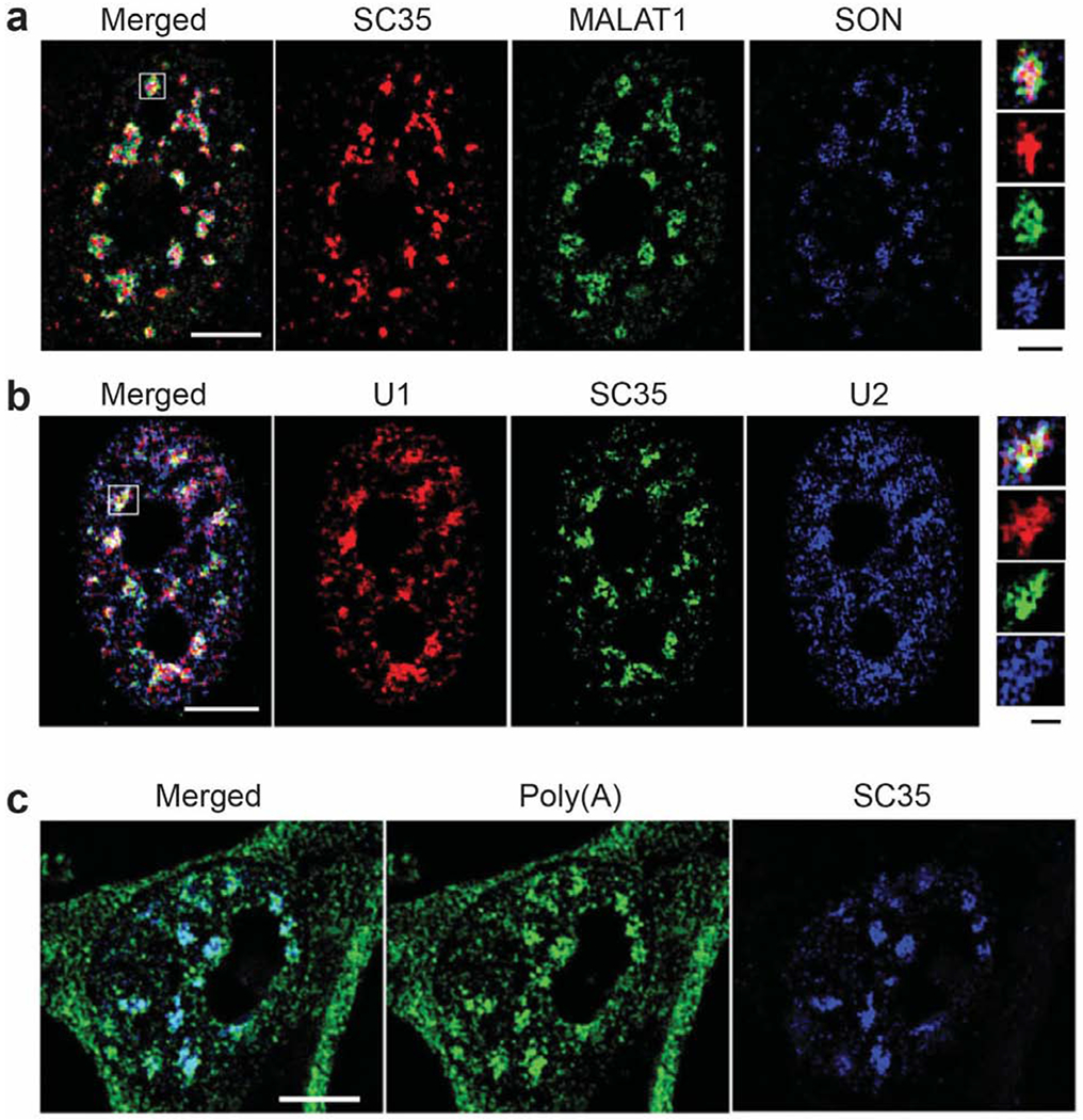
Figure 3.

Structured illumination microscopy images of nuclear speckles adapted with permission from the Journal of Cell Science, Fei et al. (2017) [50]. (a) Shown here are nuclear speckle components including the MALAT1 lncRNA, the SR-like protein SON, and many SR proteins detected by the SC35 antibody. SON and SC35 are in the nuclear speckle interior whereas MALAT1 is closer to the periphery. (b) U1 and U2 snRNAs localize to the periphery of nuclear speckles, outside of SC35. (c) Poly(A) RNA is highly enriched in nuclear speckles. White scale bars: 5 μm. Black scalebar of insets: 1 μm.
