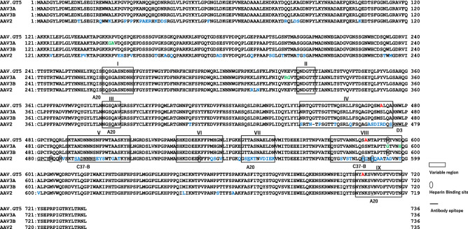
Figure 1.
Comparison of VP1 amino acids of AAV.GT5 with AAV2, AAV3A, and AAV3B. Surface variable regions (VR-1 to VR-IX) are boxed. Amino acid residues that bind to heparan sulfate are indicated with ellipse. Epitopes of antibodies against AAV2 (A20, D3, and C37-B) are underlined. Substitution of three amino acids, S472A, S587A, and N706A, on the surface loop of AAV3 are shown in red. Amino acids in AAV2 and AAV3A that are different from those in AAV3B are shown in blue and green, respectively.

