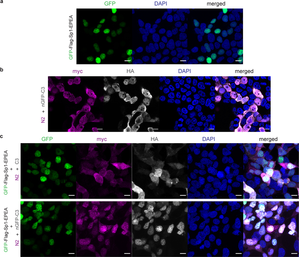
Extended Data Fig. 6. Confocal imaging of intracellular distributions of GFP-Sp1 and the split OGAs in HEK 293T cells.
a, GFP-Sp1 localized in nucleus. b, Intracellular distributions of N2 and nGFP-C3 fragments when co-expressed in HEK 293T cells. Two fragments of nGFP-splitOGA were distributed on both cytoplasm and nucleus. c, Subcellular localizations of GFP-Sp1, N fragment and C fragment when expressed simultaneously in HEK 293T cells. Two fragments of split OGA without nGFP (c, upper row) were distributed on both cytoplasm and nucleus. C-terminal fragment of nGFP-splitOGA (c, bottom row) reveals better colocalization with nuclear protein GFP-Sp1, showing the binding between nGFP and GFP. Split OGAs do not change the subcellular localization of GFP-Sp1. Channels are annotated on the top. Scale bar: 10 μm. Right: merged channel. Proteins co-expressed in each sample were labeled on the left side. Images are representative of at least three randomly selected frames.

