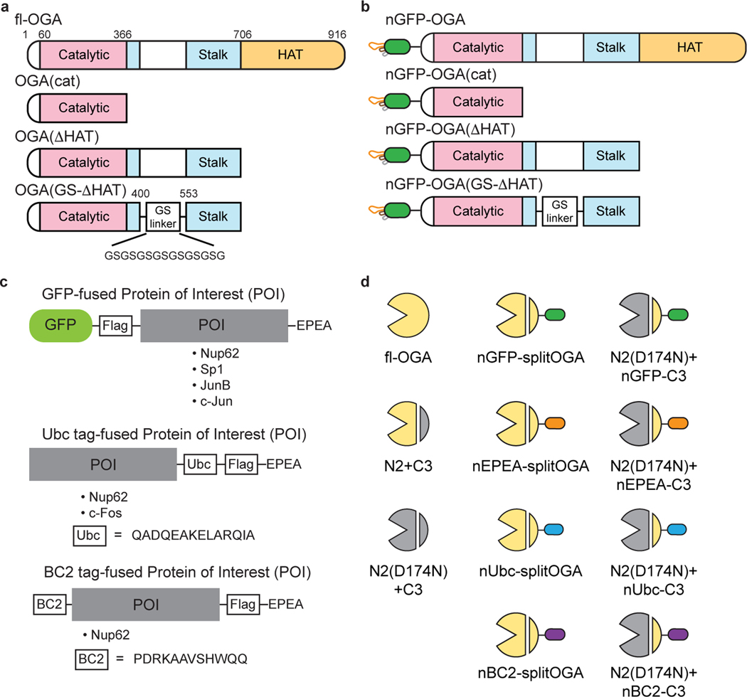
Extended Data Fig. 1. Schematic representation of OGA and target protein constructs used in this study.
a, Schematic of the structures of human OGA and other truncations. Catalytic domain, stalk domain, HAT domain, and intrinsic disordered regions are shown in pink, cyan, orange and white, respectively. GS linker represents a 15-residue glycine and serine linker. b, Depiction of the strategy to fuse the nanobody on OGAs to achieve protein specificity. nGFP, nanobody against GFP. c, Design of GFP-fused, Ubc tag-fused and BC2 tag-fused proteins of interest used in this study. For GFP-fused proteins, GFP and a Flag tag are placed on the N-terminus and the EPEA tag is in the C-terminus unless otherwise noted. For Ubc tag-fused proteins, the 14-residue peptide tag, Flag tag and EPEA tag are sequentially placed in the C-terminus unless otherwise noted. For BC2 tag-fused proteins, the 12-residue peptide tag is placed on the N-terminus and Flag, EPEA tag are in the C-terminus. Peptide sequences of Ubc and BC2 were shown. d, Symbols used in this manuscript to represent the indicated split OGA constructs.

