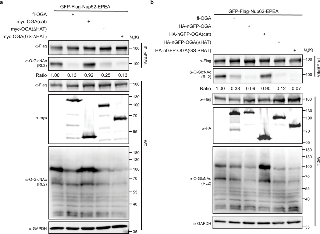
Extended Data Fig. 2. Identification of the minimal OGA for nanobody-directed deglycosylation on the target protein.
a, Enzymatic activities of OGA and its truncations are evaluated on GFP-Nup62. GFP-Nup62 was co-expressed with indicated constructs, enriched by anti-EPEA beads, and analyzed by immunoblotting to visualize the protein level and O-GlcNAc modification level, respectively. b, Evaluation of enzymatic activities of nGFP-OGA fusion proteins on GFP-Nup62. Expression levels of the indicated proteins and degree of O-GlcNAc modification were quantified by immunoblotting. The ratio equals to the intensity of anti-O-GlcNAc immunoblot normalized by the intensity of anti-Flag immunoblot. WCL, whole cell lysate. The data are representative of two biological replicates.

