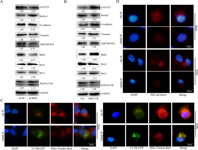
Figure 4.
BMI1 gene expression negatively correlated with autophagy of cervical cancer cells while it positively correlated with EMT. (A) HeLa cells were seeded to 6-well plates at a density of 5 × 10⁵ cells/well, transfected with shBMI1 plasmid, and protein was extracted 48 h later. BMI1, autophagy (Beclin-1, LC3IIB, Bcl-2, and P62) and EMT (N-cadherin, β-catenin, and vimentin) marker proteins as well as P-STAT3 signaling pathway were detected by western blot. (B) HeLa cells were seeded to 6-well plates at a density 5 × 10⁵ cells/well, and protein was extracted 48 h later. BMI1, autophagy (Beclin-1, LC3IIB, Bcl-2, and P62) and EMT (N-cadherin, β-catenin, and vimentin) marker proteins as well as P-STAT3 signaling pathway were detected by western blot. Cropped blots were displayed in A and B, all full-length blots are included in the Supplementary Information file. (C) HeLa and SiHa cells were seeded to 24-well plates at a density 5 × 10⁵ cells/well, transfected with shBMI1 plasmid for 24 h, infected with Ad-GFP-LC3B for 24 h, and then mitochondria were stained with Mito-Tracker Red CMXRos. Fluorescence microscope was used to observe the increase in autophagosomes and mitochondrial damage. Scale bar: 20 μm. (D) HeLa and SiHa cells were seeded to 24-well plates at a density of 5 × 10⁵ cells/well, transfected with shBMI1 plasmid for 24 h, infected with Ad-mcherry-P62 for 24 h, and then fluorescence microscope was used to observe expression of autophagy-related protein P62. Scale bar: 20 μm.*P < 0.05, vs control.

