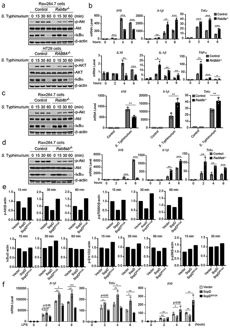
Figure 2. SopD enhances pro-inflammatory signaling by antagonizing Rab8 through its GAP activity.
(a-d) Effects of Rab8a or Rab8b deficiency on S. Typhimurium-induced Akt and NF-κB signaling. Control and Rab8a-, Rab8b-, or Rab8a/b-deficient Raw264.7 or HT29 (as indicated) cells were infected with wild-type S. Typhimurium with a multiplicity of infection of 2 and 10, respectively. At the indicated times after infection, the levels of phosphorylated AKT, the total levels of I-κBα, and the mRNA levels of the indicated cytokines were quantified by immunoblotting and qPCR. Values are the mean ± SD of three independent determinations. * P < 0.05, ** P < 0.01, *** P < 0.001, ns: not significant P > 0.05 (unpaired two-sided t test). The quantification of the immunoblots is shown in Extended Data Fig. 3. (e and f) Effect of the expression of SopD or its catalytic mutant SopDR312A on LPS-induced Akt, p70S6K, Erk1/2, p38 MAP, and NF-κB signaling and cytokine gene expression. Raw264.7 cells stably expressing HA-tagged SopD or its GAP-deficient mutant SopDR312A were treated with LPS (100 ng/ml) for the indicated times, lysed, and analyzed by immunoblotting with antibodies specific for the phosphorylated state of Akt, p70S6K, p38, and Erk1/2, as well as an antibody to I-κBα and β-actin (as a loading control). The quantification of the western blot analyses is shown (e) A repetition of this experiment is shown in Supplementary Fig. 4. Values represent the mean ± SD of three independent determinations. * P < 0.05, ** P < 0.01, *** P < 0.001, ns: not significant P > 0.05 (unpaired two-sided t test). Alternatively (f), Raw264.7 cells stably expressing HA-tagged SopD or its GAP-deficient mutant SopDR312A were treated with LPS (50 ng/ml) for the indicated times and the mRNA levels of the indicated cytokines were quantified by qPCR at the indicated times after treatment. Values are the mean ± SD of three independent determinations. * P < 0.05,** P < 0.01, *** P < 0.001, ns: not significant P > 0.05 (unpaired two-sided t test).

