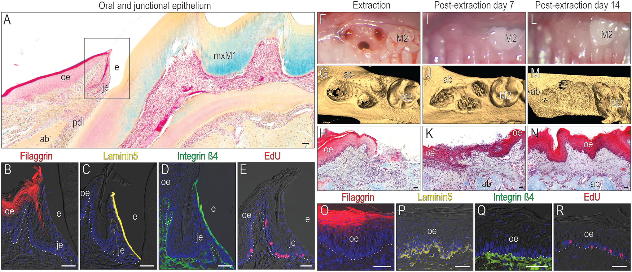
Figure 1. After tooth extraction, JE barrier characteristics are replaced with OE barrier functions.

(A) A representative tissue section stained with pentachrome to illustrate keratinized OE (red color), non-keratinized JE (pink), alveolar bone (yellow gold) and connective tissue/PDL. In the OE and JE (the black box area in A), immunohistochemical localization of (B) the terminal keratinocyte marker Filaggrin, (C) the hemidesmosomal markers Laminin5 and (D) Integrin β4; and (E) mitotically active cells that have incorporated EdU. (F) The mxM1 extraction socket viewed clinically and (G) using μCT imaging. (H) Representative tissue section stained with Masson’s trichrome from the post-extraction day (PED) 0 extraction socket. (I) Clinical, (J) μCT, and (K) histological assessment of the PED7 site. (L) Clinical, (M) μCT, and (N) histological assessment of the PED14 site. In this OE, examined 14 days after tooth extraction e.g., PED14, immunohistochemical localization of (O) Filaggrin, (P) Laminin5, and (Q) Integrin β4. (R) Mitotically active cells are identified by EdU. Dotted white lines indicate the demarcation between the epithelium and connective tissue. Abbreviations: ab, alveolar bone; e, enamel; je, junctional epithelium; oe, oral epithelium; pdl, periodontal ligament; mxM1, maxillary first molar; M2, maxillary second molar. Scale bars: 50 μm.
