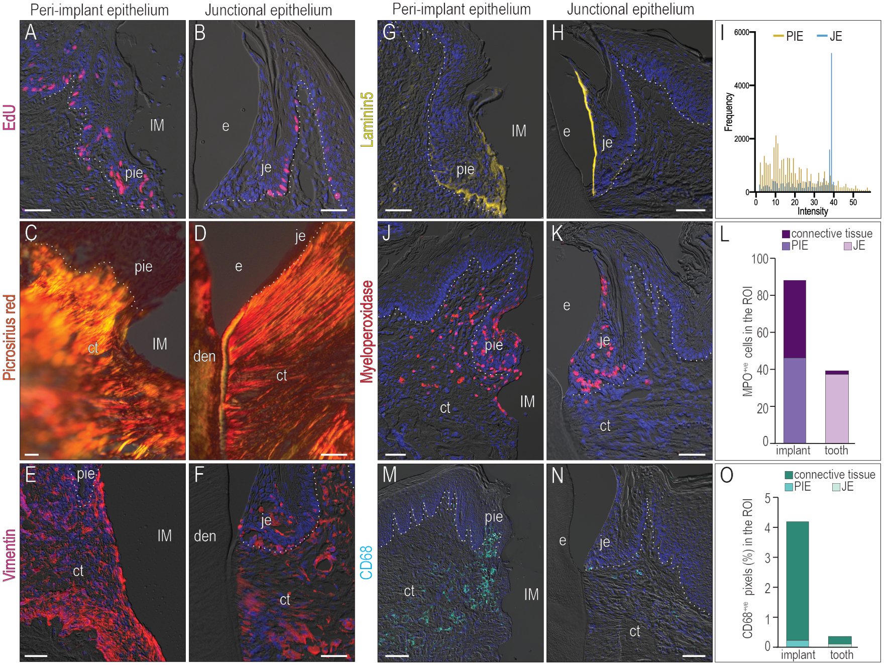
Figure 5. The PIE exhibits signs of chronic inflammation.

Mitotically active cells were labeled by EdU in (A) the PID14 PIE and (B) the JE. The fibrosis of the connective tissue was examined by (C,D) Picrosirius red staining and (E,F) Vimentin staining. The attachment to (G) the implant and (H) the tooth was evaluated by Laminin5 staining. (I) Histogram of Laminin5 expression. Immunostaining for Myeloperoxidase (MPO), a marker for neutrophils, in the (J) PIE and (K) JE. (L) Quantification of MPO+ve cells in the JE, PIE and connective tissue beneath them. Immunostaining for CD68, a marker for macrophages and monocytes, in the (M) PIE and (N) JE. (O) Quantification of CD68 expression in the JE, PIE and connective tissue beneath them. Dotted white lines indicate the demarcation between the epithelium and connective tissue. Abbreviations: as in Fig. 2 and den, dentin; ct, connective tissue.
