
Figure 1.
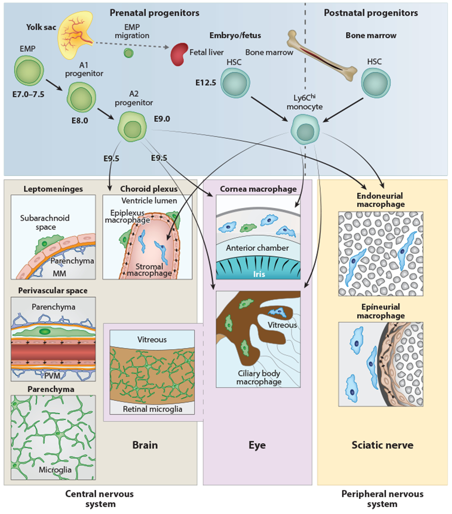
Anatomy and origin of microglia and CNS-associated macrophages. Cellular sources and anatomical location of myeloid cells found in the CNS, peripheral nervous system, or eye in the mouse. Microglial cells, PVMs, MMs, and retinal microglia are exclusively derived from prenatal sources and have no exchange with HSC-derived monocytes during postnatal stages. In contrast, choroid plexus stromal macrophages, ciliary body macrophages, corneal macrophages in the eye, endoneurial macrophages, and epineurial macrophages in the sciatic nerve have a mixed origin, from prenatal (yolk sac and fetal liver) and postnatal (bone marrow) sources. A transient early wave of myeloid cell development called primitive hematopoiesis takes place at E7.0–E7.5 (upper left). At this time, c-Kit+ EMP cells develop in the blood islands of the yolk sac. Their progeny (myeloid precursor cells) mature, expand, and migrate. CX3CR1+ A2 myeloid progenitor cells are derived from c-Kit+ CX3CR1+ A1 progenitors and seed the brain at E9.5, where they differentiate into microglial cells, PVMs, ATMs, and retinal microglia, respectively. During further development, some EMPs travel to the fetal liver (which is part of the definitive hematopoiesis), where they differentiate into monocytes. Myelopoiesis is thought to be restricted to bone marrow starting from birth (right). Abbreviations: CNS, central nervous system; EMP, erythromyeloid progenitor; HSC, hematopoietic stem cell; ATM, meningeal macrophage; PVM, perivascular macrophage.