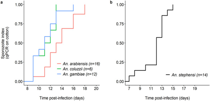
Figure 2.
The extrinsic incubation period of Plasmodium falciparum in four Anopheles mosquito species. (a) Kaplan–Meier curves representing the temporal dynamics of sporozoite appearance in small pieces of cotton used to collect saliva from individual mosquitoes in the three major African vectors An. arabiensis (red), An. gambiae (blue) and An. coluzzii (green). (b) Same as (a) but for An. stephensi. The numbers in brackets indicate the number of females for each species of mosquito that generated at least one positive cotton. African vectors were infected with the P. falciparum isolate C and An. stephensi with the NF54 laboratory culture.

