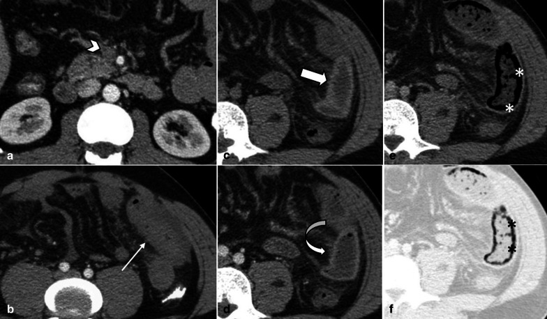
Fig. 6.
A 32 y/o male presenting with acute SMV thrombosis due to protein C deficiency. CT images on first day of admission (a, b) display thrombosis (arrowhead) in SMV with segmental circumferential mural thickening in ileum (thin arrow), mesenteric fat stranding and free fluid. On day 3 (c, d), mural hyperdensity (thick arrow) is noted in non-contrast CT at the same segment without post contrast hyper-enhancement (curved arrow) suggestive of intramural hemorrhage. Mural thickening has relatively decreased. On day 9 (e, f), mural pneumatosis (asterisks) and thinning (paper thin) are depicted consistent with development of transmural necrosis which was confirmed in laparotomy

