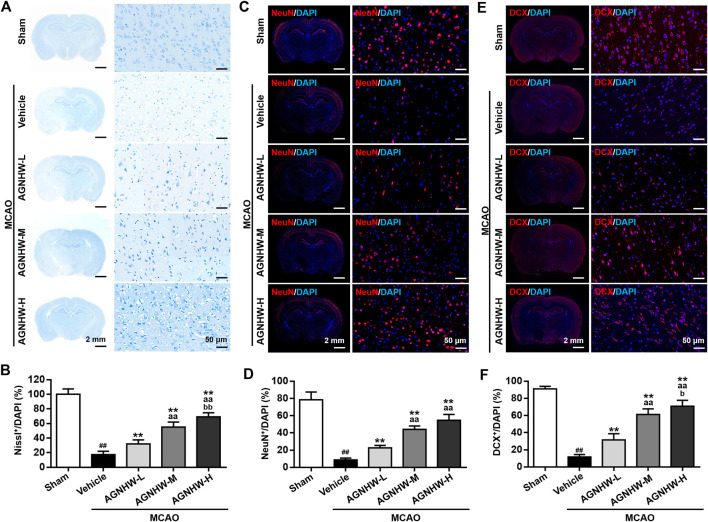
FIGURE 2.
AGNHW pre-treatment restored neuronal loss in MCAO model (A,B) Representative images (A) and statistical analysis results (B) of Nissl staining in the infarct area of the ischemic cortex in MCAO rats with or without pre-treatment with AGNHW (C,D) Representative images (C) and statistical analysis results (D) of immunofluorescent staining to visualize NeuN in the infarct area of the ischemic cortex in MCAO rats with or without pre-treatment with AGNHW (E,F) Representative images (E) and statistical analysis results (F) of immunofluorescent staining to visualize DCX in the infarct area of the ischemic cortex in MCAO rats with or without pre-treatment with AGNHW. N = 5 rats per group. Data are means ± S.D. ## p < 0.01, compared with the sham group; **p < 0.01, compared with the vehicle group; aa P<0.01, compared with AGNHW-L group; b P<0.05 and bb P<0.01, compared with the AGNHW-M group.

