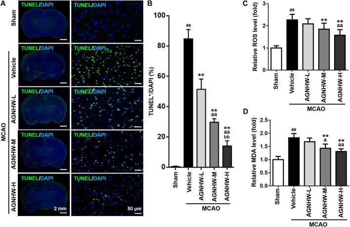
FIGURE 3.
Pre-treatment with AGNHW suppressed cell apoptosis and oxidative stress damage in MCAO model (A,B) Representative images (A) and results of statistical analysis (B) of TUNEL staining in the infarct area of the ischemic cortex in MCAO rats with or without pre-treatment with AGNHW (C, D) The status of oxidative stress was monitored by cerebral ROS (C) and MDA (D) levels, analyzed by appropriate assay kits. N = 5 rats per group. Data are means ± S.D. ## p < 0.01, compared with sham group; **p < 0.01, compared with vehicle group; a P < 0.05 and aa P<0.01, compared with AGNHW-L group; bb P<0.01, compared with AGNHW-M group.

